Epigenetics and memory

Epigenetic modification is a powerful mechanism for the induction, the expression and persistence of long-term memory.

For long-term memory, we need to consider diverse cellular processes. These occur in neurons from different brain regions (in particular hippocampus, cortex, amygdala) during memory consolidation and recall. For instance, long-term changes in kinase expression in the proteome, changes in receptor subunit composition and localization at synaptic/dendritic membranes, epigenetic modifications of chromatin such as DNA methylation and histone methylation in the nucleus, and the posttranslational modifications of histones, including phosphorylation and acetylation, all these play a role. Histone acetylation is of particular interest because a number of known medications exist, which function as histone deacetylase inhibitors (HDACs), i.e. have a potential to increase DNA transcription and memory (more on this in a later post).

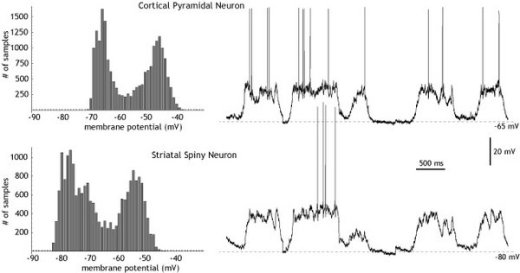
Epigenetic changes are important because they define the internal conditions for plasticity for the individual neuron. They underlie for instance, kinase or phosphatase-mediated (de)activations of enzymatic proteins and therefore influence the probability of membrane proteins to become altered by synaptic activation.

Among epigenetic changes, DNA methylation typically acts to alter, often to repress, DNA transcription at cytosine, or CpG islands in vertebrates. DNA methylation is mediated by enzymes such as Tet3, which catalyses an important step in the demethylation of DNA. In dentate gyrus of live rats, it was shown that the expression of Tet3 is greatly increased by LTP – synaptically induced memorization – , suggesting that certain DNA stretches were demethylated [5], and presumably activated. During induction of LTP by high frequency electrical stimulation, DNA methylation is changed specifically for certain genes known for their role in neural plasticity [1]. The expression of neural plasticity genes is widely correlated with the methylation status of the corresponding DNA .

So there is interesting evidence for filtering the induction of plasticity via the epigenetic landscape and modifiers of gene expression, such as HDACs. Substances which act as histone deacetylase inhibitors (HDACs) increase histone acetylation. An interesting result from research on fear suggests that HDACs increase some DNA transcription, and enhance specifically fear extinction memories [2], [3],[4]. 

Ion channel expression is not regulated by spiking behavior

An important topic to understand intrinsic excitability is the distribution and activation of ion channels. In this respect the co-regulations between ion channels are of significant interest. MacLean et al. (2003) could show that overexpression of an A-type potassium channel by shal-RNA-injection in neurons of the stomatogastric ganglion of the lobster is compensated by upregulation of Ih such that the spiking behavior remained unaltered.

A non functional shal-mutant whose overexpression did not affect spiking had the same effect, which shows that the regulation does not happen at the site of the membrane, by measuring the spiking behavior. In this case, Ih was upregulated, even though IA activity was unaltered, and spiking behavior was increased. (This is in contrast to e.g. O’Leary et al., 2013, who assume homeostatic regulation of ion channel expression at the membrane, by spiking behavior.)

In drosophila-motoneurons the expression of shal and shaker – both responsible for IA – is reciprocally coupled. If one is reduced, the other is upregulated to a constant level of IA activity at the membrane. Other ion channels, like (INAp and IM) are again antagonistic, which means they correlate positively: if one is reduced, the other is reduced as well to achieve the same level of effect (Golowasch2014). There are a number of publications which have all documented similar effects, e.g. (MacLean et al., 2005, Schulz et al., 2007; Tobin et al., 2009; O’Leary et al., 2013).

We must assume that the expression level of ion channels is regulated and sensed inside the cell and that the levels of genes for different ion channels are coupled – by genetic regulation or on the level of RNA regulation.

To summarize: When there is high IA expression, Ih is also upregulated. When one gene responsible for IA is suppressed, the other gene is more highly expressed, to achieve the same level of IA expression. When (INap), a permanent sodium channel, is reduced, (IM), a potassium channel, is also reduced.

It is important to note that these ion channels may compensate for each other in terms of overall spiking behavior, but they have subtly different properties of activation, e.g. by the pattern of spiking or by neuromodulation. For instance, if cell A reduces ion channel currents like INap and IM, compensating to achieve the same spiking behavior, once we apply neuromodulation to muscarinic receptors on A, this will affect IM, but not INap. The behavior of cell A, crudely the same, is now altered under certain conditions.

To model this – other than by a full internal cell model – requires internal state variables which guide ion channel expression, and therefore regulate intrinsic excitability. These variables would model ion channel proteins and their respective interaction, and in this way guarantee acceptable spiking behavior of the cell. This could lead to the idea of an internal module which sets the parameters necessary for the neuron to function. Such an internal module that self-organizes its state variables according to specified objective functions could greatly simplify systems design. Instead of tuning systems parameters by outside methods – which is necessary for ion-channel based models – each neuronal unit itself would be responsible for its ion channels and be able to self-tune them separately from the whole system.

Linked to the idea of internal state variables is the idea of internal memory, which I have referred to several times in this blog. If I have an internal module of co-regulated variables, which set external parameters for each unit, then this module may serve as a latent memory for variables which are not expressed at the membrane at the present time (s. Er81). The time course of expression and activation at the membrane and of internal co-regulation need not be the same. This offers an opportunity for memory inside the cell, separated from information processing within a network of neurons.

Egocentric representations for general input

The individual neuron’s state need not be determined only by the inputs received.

(a). It may additionally be seeded with a probability for adaptation that is distributed wrt the graph properties of the neuron (like betweenness centrality, choke points etc.), as well as the neuron’s current intrinsic excitability (IE) (which are related). This seeded probability would correspond to a sensitivity of the neuron to the representation that is produced by the subnetwork. The input representation is transformed by the properties of the subnetwork.

(b). Another way to influence neurons independent of their input is to link them together. This can be done by simulating of neuromodulators (NMs) which influence adaptivity for a subset of neurons within the network. There are then neurons which are linked together and increase or turn on their adaptivity because they share the same NM receptors. Different sets of neurons can become activated and increase their adaptivity, whenever a sufficient level of a NM is reached. An additional learning task is then to identify suitable sets of neurons. For instance, neurons may encode aspects of the input representation that result from additional, i.e. attentional, signals co-occuring with the input.

(c). Finally, both E and I neurons are known to consist of morphologically and genetically distinct types. This opens up additional ways of creating heterogeneous networks from these neuron types and have distinct adaptation rules for them. Some of the neurons may not even be adaptive, or barely adaptive, while others may be adaptive only once, (write once, read-only), or be capable only of upregulation, until they have received their limit. (This applies to synaptic and intrinsic adaptation). Certain neurons may have to follow the idea of unlimited adaptation in both directions in order to make such models viable.

Similar variants in neuron behavior are known from technical applications of ANNs: hyperparameters that link individual parameters into groups (‘weight sharing’) have been used, terms like ‘bypassing’ mean that some neurons do not adjust, only transmit, and ‘gating’ means that neurons may regulate the extent of transmission of a signal (cf. LSTM, ScardapaneSetal2018). Separately, the model ADAM (or ADAMW) has been proposed which computes adaptive learning rates for each neuron and achieves fast convergence.

A neuron-centric biological network model (‘neuronal automaton’) offers a systematic approach to such differences in adaptation. As suggested, biological neurons have different capacities for adaptation and this may extend to their synaptic connections as well. The model would allow to learn different activation functions and different adaptivity for each neuron, helped by linking neurons into groups and using fixed genetic types in the setup of the network. In each specific case the input is represented by the structural and functional constraints of the network and therefore transformed into an internal, egocentric representation.

Antagonistic regulation for cellular intelligence

Cellular intelligence refers to information processing in single cells, i.e. genetic regulation, protein signaling and metabolic processing, all tightly integrated with each other. The goal is to uncover general ‘rules of life’ wrt e.g. the transmission of information, homeostatic and multistable regulation, learning and memory (habituation, sensitization etc.). These principles extend from unicellular organisms like bacteria to specialized cells, which are parts of a multicellular organism.

A prominent example is the ubiquitous role of feedback cycles in cellular information processing. These are often nested, or connected to a central hub, as a set of negative feedback cycles, sometimes interspersed with positive feedback cycles as well. Starting from Norbert Wiener’s work on cybernetics, we have gained a deeper understanding of this regulatory motif, and the complex modules that can be built from a multitude of these cycles by modeling as well as mathematical analysis.

Another motif that is similar in significance and ubiquity is antagonistic interaction. A prototypical antagonistic interaction consists of a signal, two pathways, one negative, one positive, and a target. The signal connects to the target by both pathways. No further parts are required.

On the face of it, this interaction seems redundant. When you connect a signal to a target by a positive and a negative connection, the amount of change is a sum of both connections, and for this, one connection should be sufficient. But this motif is actually very widespread and powerful, and there are two main aspects to this:

A. Gearshifting, scale-invariance or digitalization of input: for an input signal that can occur at different strengths, the antagonistic transmission allows to shift the signal to a lower level/gear with a limited bandwidth compared to the input range. This can also be described as scale-invariance or standardization of the input, or in the extreme case, digitalization of an analog input signal.

B. Fast onset-slow offset response curves: in this case the double transmission lines are used with a time delay. The positive interaction is fast, the negative interaction is slow. Therefore there is a fast peak response with a slower relaxation time– useful in many biological contexts, where fast reaction times are crucial.

Negative feedback cycles which can achieve similar effects by acting on the signal itself: the positive signal is counteracted by a negative input which reduces the input signal. The result is again a fast peak response followed by a downregulation to an equilibrium value. The advantage for antagonistic interactions is that the original signal is left intact, which is useful. because the same signal may act on other targets unchanged. In a feedback cycle the signal itself is consumed by the feedback interaction. The characteristic shape of the signal, fast peak response with a slower downregulation, may therefore arise from different structures.

The type of modules that can be built from both antagonistic interactions and feedback have not been explored systematically. However, one example is morphogenetic patterning, often referred to as ‘Turing patterns’, which relies on a positive feedback cycle by an activator, plus antagonistic interactions (activator/inhibitor) with a time delay for the inhibitor.

 turing_pattern

Transmission is not Adaptation

Current synaptic plasticity models have one decisive property which may not be biologically adequate, and which has important repercussions on the type of memory and learning algorithms in general that can be implemented: Each processing or transmission event is an adaptive learning event.

In contrast, in biology, there are many pathways that may act as filters from the use of a synapse to the adaptation of its strength. In LTP/LTD, typically 20 minutes are necessary to observe the effects. This requires the activation of intracellular pathways, often co-occurence of a GPCR activation, and even nuclear read-out.

Therefore we have suggested a different model, greatly simplified at first to test its algorithmic properties. We include intrinsic excitability in learning (LTP-IE, LTD-IE). The main innovation is that we separate learning or adaptation from processing or transmission. Transmission events leave traces at synapses and neurons that disappear over time (short-term plasticity), unless they add up over time to unusually high (low) neural activations, something that can be determined by threshold parameters. Only if a neuron engages in a high (low) activation-plasticity event we get long-term plasticity at both neurons and synapses, in a localized way. Such a model is in principle capable of operating in a sandpile fashion. We do not know yet what properties the model may exhibit. Certain hypotheses exist, concerning abstraction and compression of a sequence of related inputs, and the development of an individual knowledge.

Neuromodulation and Cortical Computation

The impact of neuromodulation on cortical computation is still an area of active debate. Recently, fluctuations in synchronization under neuromodulation in cortical areas have been observed. This is important because synchronization in a network influences the read-out of intrinsic membrane properties, i.e. neural excitability. Synchronization shifts a neuron from transmission mode (strong correlation) to intrinsic mode (weak correlation). Here we show how neuromodulation through its impact on synapses may alter the topology of network connections, and how this leads to the observed neuromodulation-dependent fluctuations in synchronization. In our theory, neuromodulation is vitally important for computation, because it is selective for cortical areas, influences the degree of internal synchronization, and therefore defines the actual state of information-processing in the area.

via Neuromodulation Influences Synchronization and Intrinsic Read-outdrugs-medicinal-omega-3-grass-48194.jpeg

Consciousness made easy

IMS Photo Contest 2017

For a long time I didn’t know what research on consciousness was to be about. Was it being able to feel and think? Was it perceptual awareness (as in ‘did you hear that sound’?) What did attention have to do with it (the searchlight hypothesis), i.e. lots of stored information is present but not ‘in consciousness’ at any given moment in time?
Finally, while discussing  the issue that no one has a good theory of anesthesia (TMK), (i.e. how it happens and why it works), it occurred to me we can simplify the question, and make it solvable in a fairly easy way:
Consciousness (C) made easy is just the difference between awake state W  and anesthesia/slow wave sleep SWS/A.

C = W – SWS/A

The difference is what makes up consciousness. We can measure this difference in a number of ways, brain imaging, neuronal spiking behavior, EEG/EcoG, LFPs, voltammetry of neurochemicals, possibly gene expression, and quantify it. Sure it is not a simple task, and people may disagree on how to integrate measurements for a solid theory of what is happening, but conceptually it is at least clearly defined.

600px-CjwUpDownStateFig1

Charles Wilson (2008), Scholarpedia, 3(6):1410.               doi:10.4249/scholarpedia.1410

An important difference is the appearance of up-and down states when unconscious. Possibly in this state only the purely mechanical coupling of the neuronal mass remains, and the fine-tuned interactions by chemical receptors and channels is simplified such that the high entropy asynchronous spiking is abolished.

It would be interesting to further investigate the soliton theory for this question.


Memory and the Volatility of Spines

Memory has a physical presence in the brain, but there are no elements which permanently code for it.

Memory is located – among other places – in dendritic spines. Spines are being increased during learning and they carry stimulus or task-specific information. Ablation of spines destroys this information (Hayashi-Takagi A2015). Astrocytes have filopodia which are also extended and retracted and make contact with neuronal synapses. The presence of memory in the spine fits to a neuron-centric view: Spine protrusion and retraction are guided by cellular programs. A strict causality such that x synaptic inputs cause a new spine is not necessarily true, as a matter of fact highly conditional principles of spine formation or dissolution could hold, where the internal state of the neuron and the neuron’s history matters. The rules for spine formation need not be identical to the rules for synapse formation and weight updating (which depend on at least two neurons making contact).

A spine needs to be there for a synapse to exist (in spiny neurons), but once it is there, clearly not all synapses are alike. They differ in the amount of AMPA presence and integration, and other receptors/ion channels as well. For instance, Sk-channels serve to block off a synapse from further change, and may be regarded as a form of overwrite protection. Therefore, the existence or lack of a spine is the first-order adaptation in a spiny neuron, the second-order adaptation involves the synapses themselves.

However, spines are also subject to high variability, on the order of several hours to a few days. Some elements may have very long persistence, months in the mouse, but they are few. MongilloGetal2017 point out the fragility of the synapse and the dendritic spine in pyramidal neurons and ask what this means for the physical basis of memory. Given what we know about neural networks, for memory to be permanent, is it necessary that the same spines remain? Learning allows to operate with many random elements, but memory has prima facie no need for volatility.

It is most likely that memory is a secondary, ’emergent’ property of volatile and highly adaptive structures. From this perspective it is sufficient to keep the information alive, among the information-carrying units, which will recreate it in some form.

The argument is that the information is redundantly coded. So if part of the coding is missing, the rest still carries enough information to inform the system, which recruits new parts to carry the information. The information is never lost, because not all synapses, spines, neurons are degraded at the same time, and because internal reentrant processing keeps the information alive and recreates new redundant parts at the same time as other parts are lost. It is a dynamic cycling of information. There are difficulties, if synapses are supposed to carry the whole information. The main difficulty is: if all patterns at all times are being stored in synaptic values, without undue interference, and with all the complex processes of memory, forgetting, retrieval, reconsolidation etc., can this be fitted to a situation, where the response to a simple visual stimulus already involves 30-40% of the cortical area where there is processing going on? I have no quantitative model for this. I think the model only works if we use all the multiple, redundant forms of plasticity that the neuron possesses: internal states, intrinsic properties, synaptic and morphological properties, axonal growth, presynaptic plasticity.

Balanced Inhibition/Excitation (2) – The I/E ratio

Some time ago, I suggested that the theoretical view on balanced inhibition/excitation (in cortex and cortical models) is probably flawed. I suggested that we have a loose regulation instead, where inhibition and excitation can fluctuate independently.

The I/E balance stems from the idea that the single pyramidal neuron should receive approximately equal strength of inhibition and excitation, in spite of the fact that only 10-20% of neurons in cortex are inhibitory (Destexhe2003, more on that below). Experimental measurements have shown that this conjecture is approximately correct, i.e. inhibitory neurons make stronger contacts, or their influence is stronger relative to excitatory inputs.

The E-I balance in terms of synaptic drive onto a single pyramidal neuron is an instance of antagonistic regulation which allows gear-shifting of inputs, and in this case, allows very strong inputs to be downshifted by inhibition to a weaker effect on the membrane potential. What is the advantage of such a scheme? Strong signals are less prone to noise and uncertainty than weak signals. Weak signals are filtered out by the inhibitory drive. Strong signals allow unequivocal signal transmission, whether excitatory synaptic input, (or phasic increases of dopamine levels, in other contexts), which are then gear-shifted down by antagonistic reception. There may also be a temporal sequence: a strong signal is followed by a negative signal to restrict its time course and reduce impact. In the case of somatic inhibition following dendritic excitation the fine temporal structure could work together with the antagonistic gear-shifting exactly for this goal. Okun and Lampl, 2008 have actually shown that inhibition follows excitation by several milliseconds.

But what are the implications for an E/I network, such as cortex?

Here is an experimental result:

During both task and delay, mediodorsal thalamic (MD) neurons have 30-50% raised firing rates, fast-spiking (FS) inhibitory cortical neurons have likewise 40-60% raised firing rates, but excitatory (regular-spiking, RS) cortical neurons are unaltered. Thus there is an intervention possible, by external input from MD, probably directly to FS neurons, which does not affect RS neuron rate at all (fig. a and c, SchmittLIetal2017)

Untitled 1

Mediodorsal thalamic stimulation raises inhibition, but leaves excitation unchanged.

At the same time, in this experiment, the E-E connectivity is raised (probably by some form of short-term synaptic potentiation), such that E neurons receive more input, which is counteracted by more inhibition. (cf. also Hamilton, L2013). The balance on the level of the single neuron would be kept, but the network exhibits only loose regulation of the I/E ratio: unilateral increase of inhibition.

There are several studies which show that it is possible to raise inhibition and thus enhance cognition, for instance in the mPFC of CNTNAP2 (neurexin, a cell adhesion protein) deficient mice, which have abnormally raised excitation, and altered social behavior (SelimbeyogluAetal2017, cf. Foss-FeigJ2017 for an overview). Also, inhibition is necessary to allow critical period learning – which is hypothesized to be due to a switch from internally generated spontaneous activity to external sensory perception (ToyoizumiT2013) – in line with our suggestion that the gear-shifting effect of locally balanced I/E allows only strong signals to drive excitation and spiking and filters weak, internally generated signals.


Some thoughts about Language Evolution

Judging by ontogenetic development, language is derived from two separate streams (and a third one later on).
One of these is the development of object and action concepts by visuomotoric handling as opposed to background, space or situation. This may well be specific to humans with infants developing eye-hand-coordination in contrast to other species which do not undergo such a phase and may not develop strong concepts for objects. Infants also form a concept for actions when they experience their own agency and extend it to other agents.
The other is the sound-making ability, which appears innately pleasurable to human infants, and contributes to a long period of babbling.
At around 10-12 months of age these developments combine (‘naming insight’ in child language literature) and articulated words which refer to simple concepts arise. These refer to objects at first, sometimes actions and are articulated by phonological sequences. The one and two word (‘pivot grammar’) stages follow.
I see no reason to assume that phylogenetic development should have been different – the development of articulatory abilities, considered beautiful for their own sake (‘music/song’) running in parallel with solid conceptual structuring of the environment before one and two word communication became commonplace.
What about communication and communicative needs? I believe they popped up after the first concept naming skills took root. Suddenly there must have been a drift towards higher information content messages rather than just a string of words denoting objects and actions. Communication then is what must have driven grammar. Note that grammar remains a highly social accomplishment – like phonological sophistication it even requires a critical period. Grammar like phonology probably has a strong striatal component – habit learning – and it spontaneously appears in sign language as well. For this we would require a more detailed theory on how grammar arose from communication, when each grammatical system is distinct from any other.